Events
Meet us during the following events
Webinar
2022/06/29
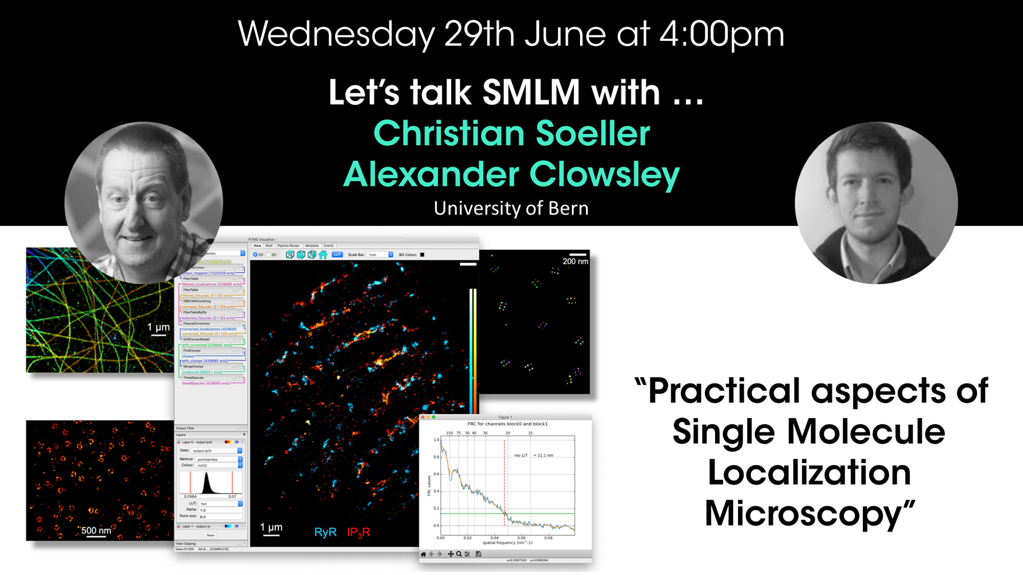
Prof. Christian Soeller and Dr Alexander Clowsley - Practical aspects of Single Molecule Localization Microscopy
Christian Soeller and Dr Alexander Clowsley
University of Bern, Switzerland
CLICK HERE TO REGISTER
Learn more
Webinar
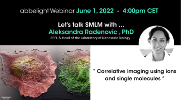
2022/06/01
Aleksandra Radenovic - Correlative imaging using ions and single molecules
Tijana Talisma
City of Hope, Comprehensive Cancer Center, CA, USA
CLICK HERE TO REGISTER
Learn more
Webinar
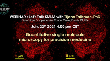
2021/07/22
Tijana Talisman - Quantitative single molecule localization microscopy for precision medicine
Tijana Talisma
City of Hope, Comprehensive Cancer Center, CA, USA
CLICK HERE TO REGISTER
Learn more
Webinar
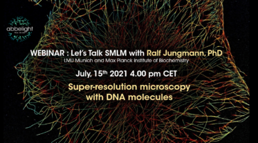
2021/07/15
Dr Ralf Jungmann - Super-resolution microscopy with DNA molecules
Ralf Jungmann
Max Planck Institute of Biochemistry, Munich
CLICK HERE TO REGISTER
Learn more
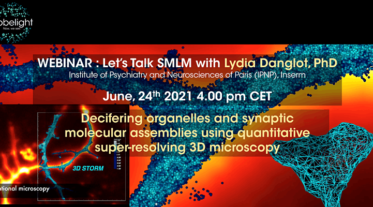
Webinar
2021/06/24
Lydia Danglot - Decifering organelles and synaptic molecular assemblies using quantitative super-resolving 3D microscopy
Lydia Danglot
Institut de Psychiatrie et Neurosciences de Paris (IPNP)
CLICK HERE TO REGISTER
Learn more
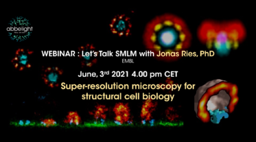
Webinar
2021/06/03
Dr. Jonas Ries - Super-resolution microscopy for structural cell biology
Jonas Ries, PhD
EMBL, Heidelberg
CLICK HERE TO REGISTER
Learn more
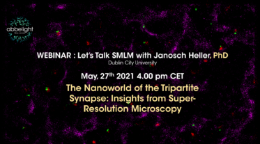
Webinar
2021/05/27
Dr. Janosch HELLER - The Nanoworld of the Tripartite Synapse
Janosch HELLER, PhD
Dublin City University
CLICK HERE TO REGISTER
Learn more
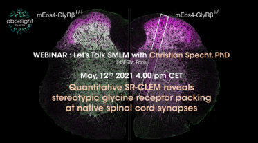
Webinar
2021/05/12
Dr Christian Specht - Quantitative SR-CLEM reveals stereotypic glycine receptor packing at native spinal cord synapses
Christian Specht, PhD
INSERM, Paris
CLICK HERE TO REGISTER
Learn more
Webinar
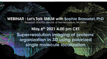
2021/05/06
Dr. Sophie Brasselet - Super resolution imaging of proteins’ organization
Sophie Brasselet, PhD
Fresnel Institute, Marseille
CLICK HERE TO REGISTER
Learn more
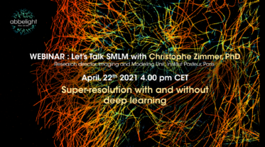
Webinar
2021/04/22
Dr. Christophe ZIMMER - SMLM with and without deep learning
Christophe Zimmer, PhD
Institut Pasteur, Paris
CLICK HERE TO REGISTER
Learn more
Event
2020/10/28
NeuroscienceWeek2020
Online
During the NeuroscienceWeek2020 organized by Olympus, we will be present to show you how single-molecule localization microscopy can be a very relevant technique for biological neuroscience research.
On Wednesday, 28th October from 10:00 to 11:30 am (EST), we propose you to participate at Abbelight talks of the Neuroscience week, with the following program with a Tech Talk on membrane compartmentalization by the neuronal membrane cytoskeleton (10:00am – 10:20am), Product Introduction of abbelight nanoscopy systems for single molecule localization microscopy (10:20am – 11:00am), Virtual demo of Abbelight’s SAFe nanoscopy systems (11:00am – 12:00pm)
Learn more
Webinar
2020/07/10
All-optical imaging of molecules in their nanoscale cellular context
Pr. Joerg Bewersdorf
Yale University School of Medicine
Super-resolution microscopy has become a powerful tool to study the nanoscale spatial distribution of proteins of interest in cells over the last years. Imaging any of these distributions in the context of other proteins or the general cellular context is, however, still challenging. In this webinar, I will present recent developments of our lab which offer a diverse set of solutions that tackle this challenge: ‘Salvaged fluorescence’ enables ratiometric 3-color single-molecule super-resolution imaging at 5–10-nm localization precision in all color channels at negligible chromatic shift or cross-talk [1]. A new fluorogenic DNA-PAINT probe enables fast, 3D whole-cell imaging without the need for optical sectioning, adding a versatile tool to the toolbox of single-molecule super-resolution probes [2]. Labeling proteins and other cellular components in bulk in our novel pan-Expansion Microscopy method provides ultrastructural context to the nanoscale organization of proteins,...
Learn more
Webinar
2020/07/03
Application of super-resolution imaging methods for cell biology and translational medicine
Dr. Vito Mennella
NIHR Biomedical Research Center, University of Southampton
Prof. Vito Mennella has pioneered the application of multimodal super-resolution imaging in cell biology, in particular to study organelle architecture (Trends in Cell Biology, 2014, 2015).
By leveraging the power of 3D-SIM, STORM microscopy and quantitative image analysis, he has made together with his team a series of paradigm-shifting discoveries in centrosome and cilia biology: 1. The organized molecular architecture of the pericentriolar material of centrosomes (Nature Cell Biology, 2012); The architecture and function of a novel centrosomal complex in situ (Elife, 2018) and characterization of a novel type of cilium in the airway (Developmental Cell, 2020, 2020 in press).
Most recently he collaborated with clinicians to build super-resolution based tools for translational research in diseases caused by motile cilia defects and apply machine learning methods for disease diagnosis (Science Translational Medicine, 2020). In his presentation he will give an overview of...
Learn more
Webinar
2020/06/26 - 4.00pm CEST
Present, future and past of super-resolution microscopy by dSTORM
Pr. Markus Sauer
Department of Biotechnology and Biophysics, Biocenter, Julius Maximilian University
One of the fathers of localization microscopy, and the inventor of dSTORM, Pr. Markus Sauer will talk about the present, the future and the past of super-resolution microscopy by dSTORM. He will briefly introduce basic requirements of localization microscopy, its potential use for quantitative molecular imaging, and discuss present obstacles and ways to bypass them. He will demonstrate the advantageous use of single-molecule localization microscopy by dSTORM for quantitative imaging of plasma membrane receptors and the molecular architecture of multiprotein complexes including imaging by 3D lattice-light-sheet dSTORM.
Learn more
Webinar
2020/06/19 - 4:00 pm CEST
Extracting biologically relevant, quantitative information from SMLM data with applications in immune cells
Dr. DYLAN OWEN
University of Birmingham
While single-molecule localisation microscopy (SMLM) can generate images of biological samples with nanometer resolution, challenges still remain in extracting relevant and quantitative information from the acquired data sets. Pr. Dylan Owen will overview his lab's work on developing a variety of software tools for describing biological structures from SMLM data and show how SMLM can answer biological questions without needing to generate images. The methods will be illustrated in immune cells where we seek to understand the role protein spatial organisation plays in the regulation of the T cell immunological synapse.
Learn more
Webinar
2020/06/12 - 4:00 pm CEST
The inner life of integrin adhesion sites, from single molecules to functional macromolecular complexes
Dr Gregory Giannone & Dr Olivier Rossier
Bordeaux University - Spatio-temporal and mechanical control of motile structures
Using recent advances in super-resolution microscopy and single protein tracking, it is now possible to localize single proteins in three dimensions inside integrin adhesions, determine their diffusive behaviors, and mechanical responses during cell stretching. Here, we will present how we have used and developed tools to link the molecular behavior of integrin adhesion proteins with their functions during integrin activation and mechano-sensing.
Learn more
Webinar
2020/06/05 - 4:00pm CEST
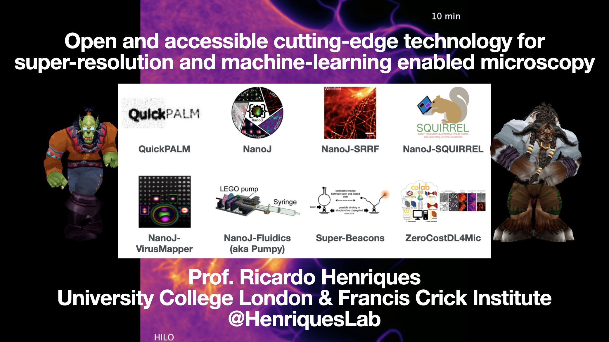
Open and accessible cutting-edge technology for super-resolution and machine-learning enabled microscopy
Pr. RICARDO HENRIQUES
University College London & Francis Crick Institute
Here I will present an overview of some of our research focused on creating open-technology to enable new observations in nanoscale live cellular imaging. All these methods are transparent, reproducible and widely available to researchers
Learn more
Webinar
2020/05/20 - 4:00 pm CEST
From single cell imaging to in vivo single-molecule biochemistry
Pr. Ulrike Endesfelder
Max Planck Institute for Terrestrial Microbiology and LOWE Center for Synthetic Microbiology (SYNMIKRO), Marburg, Germany
You should watch this webinar if you are interested in a beginners guide on how to vamp up your wide field fluorescence microscope and sample preparations to single-molecule sensitivity, including many tips and tricks from my groups work. I will explain tools such as quantitative dual-color PALM imaging using dual fluorescent protein labeling in living cells, details of sample preparations like easy but precise drift correction by red-shifted beads or tracking of dense, highly dynamic single-molecule data.
Learn more
Webinar
2020/05/15 - 4:00 pm CEST
Using super-resolution microscopy and Affimers to determine protein organization in cells with nanometre precision
Pr. Michelle Peckham
The Astbury Centre for Structural Molecular Biology, University of Leeds
I'll be talking about our use of Affimers, small non-antibody binding proteins, in confocal and super-resolution imaging and how we look for patterns in super-resolution images.
Learn more

Event
2020/04/05 > 2020/04/08
FOM 2020
Osaka
Key subjects for the conference series are the theory and practice of 3D optical imaging, related 3D image processing, and reporting especially on developments in resolution and imaging modalities. The conference series covers also the rapidly advancing fluorescence labeling techniques for confocal and multi-photon 3D imaging of -live- biological specimens.
Typical topics of the upcoming FOM conference will include:
• Theory and practice of confocal and multiphoton-excitation microscopy • Super-resolution, nanoscopy imaging: from PSF engineering (4pi, SIM, STED), fluorescent activation/quenching, stochastic/centroid (PALM, STORM, GSDIM, SOFI and related techniques) to TIRF • 3D and 4D live cell and tissue imaging • Adaptive optics for microscopy • Light sheet microscopy • Phase/interference based microscopies, • OCT, holographic, endoscopy • Advanced fluorescence imaging/spectroscopy: FRET, FRAP, FLIM, FCS, SOFI • New fluorescence probes, proteins, quantum...
Learn more
Event
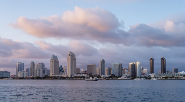
2020/02/15 > 2020/02/19
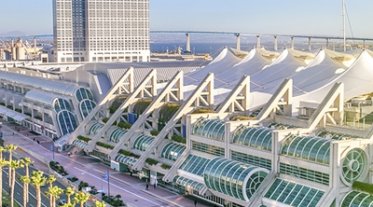
BPS 2020
San Diego, USA
As science becomes increasingly interdisciplinary, the Biophysical Society Annual Meeting continues its long-held reputation for bringing together leading scientists from the all over the world who work at the interface of the life, physical, and computational sciences.
The dynamic five-day Meeting provides attendees with opportunities to share their latest unpublished findings and learn the newest emerging techniques and applications.
Despite its nearly 6,500 attendees, the Meeting is noted for maintaining its “small meeting” feel beginning with the Saturday subgroup symposia, which allow attendees to meet within their scientific communities. It is also known for its vitality, demonstrated by the over 900 highly interactive daily poster presentations, the more than 500 speakers selected from submitted abstracts, the many career development programs for those working in academia, industry, and agencies throughout the world, and its advocacy and education programs.
Learn more
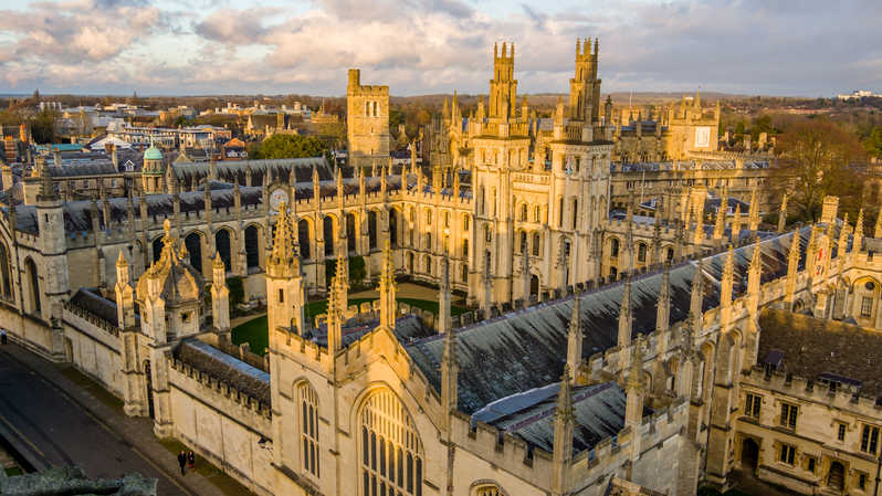
Event
2020/01/06 > 2020/01/09
QBI 2020
Oxford, UK
The QBI 2020 Conference will be held at the University of Oxford’s Mathematical Institute, Oxford, UK between January 6-9, 2020.
The idea for a conference on Quantitative BioImaging followed from the recognition that there is no conference to date that addresses, in a focused and interdisciplinary manner, the analysis of bioimaging data.
Learn more
Event
2019/12/07 > 2019/12/11
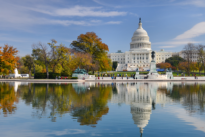
ASCB 2019
Washington DC, USA
The 2019 joint meeting of the American Society for Cell Biology (ASCB) and European Molecular Biology Organization (EMBO) will focus on cell biology as the fundamental basis of biology, and will offer sessions on emerging topics such as nontraditional model organisms, and the use of computational modeling and biophysics to “Build the Cell from the Ground Up.”
Learn more

Event
2019/09/11 > 2019/09/13
Microscopy at the Frontiers of Science 2019
The objective of the "MICROSCOPY AT THE FRONTIERS OF SCIENCE 2019" is to present and discuss the state of art and the potential of Microscopy and Microanalysis techniques in research areas such as materials and life sciences, biomaterials, geology, heritage and archaeology, forensic sciences and industrial quality and innovation.
Event
2019/08/19 > 2019/08/29
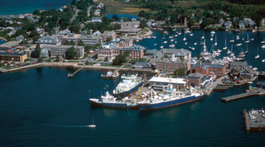
OMIBS optical microscopy course at the MBL
Woods Hole, USA
This course is designed primarily for research scientists, postdoctoral trainees, core facility directors/staff and graduate students working in the biological sciences. Biologists and physicists alike seeking a comprehensive introduction to microscopy and digital imaging will benefit greatly from the course. This 9-day course is limited to 24 students to ensure a truly interactive, hands-on experience. It consists of interrelated lectures, laboratory exercises, demonstrations, and discussions that will enable the participants to obtain and interpret high quality microscope data, to understand and assess potential artifacts, to perform quantitative optical measurements, and to generate digital images for documentation and analysis that accurately present the data. The course also places a strong emphasis on appropriate sample preparation, including choice of fluorescent probes and fluorescent proteins, and tissue clearing and refractive index matching. Particular emphasis will be...
Learn more

Event
2019/07/07 > 2019/07/11
FEMS 2019
Glasgow, Scotland
FEMS - FEDERATION OF EUROPEAN MICROBIOLOGICAL SOCIETIES
We are FEMS, a pan-European scientific body campaigning to get science - and microbiology in particular - on the European agenda. Set up in 1974 to promote microbiology across Europe, today we are a growing coalition of 53 Member Societies from 38 countries. We bring together around 30,000 professional microbiologists who are exploring microbiology and working to translate that knowledge into impact on the ground, and we are working with similar organizations elsewhere to make our network global. We publish five microbiology journals and organize a biennial congress for microbiologists around the world. Our community can apply for fellowships, grants and/or support when organizing or attending meetings and we work together with these and other partners to track, promote and recognize excellence in microbiology.
Learn more
Event
2019/07/02 > 2019/07/05
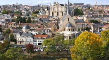
Société française des microscopies
Poitiers, France
The Sfµ meeting aims at bringing together a wide community of scientists who develop or make use of electron, photon, scanning probe and atom probe microscopies. The scientific program consists of twelve symposia: four dedicated to life science, four focused on materials science, and four joint symposia (life and materials science). This congress is synchronized with the spring meeting of GN-MEBA, providing the opportunity for shared discussions.
Learn more
Event
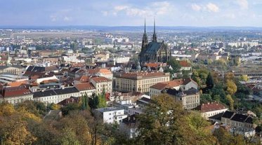
2019/06/04 > 2019/06/07
ELMI 2019
Brno, Czech Republic
ELMI conferences were established as a unique networking opportunity providing exchange of information between European scientists working in the field of light microscopy and the manufacturers of microscopy equipment. They have become a regular meeting place of scientists who develop and apply advanced light microscopy in their research, as well as becoming a crucial conference for imaging networks and people working in imaging facilities.
Learn more
Event
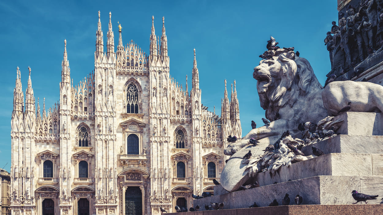
2019/06/03 > 2019/06/06
CellMech 2019
Milan, Italy
Cell Mech is a conference that has been organised every other year in Europe for the last 15 years. The conference series gathers biologists, mathematicians, physicists and engineers to discuss various aspects of cell mechanics. It has a strong focus on fostering interactions between students and established PIs in the field. The workshop format offers ample time for interactions and presentation of new or unpublished work.
This year will bring together people interested in biological forces from different origins (osmotic forces, cytoskeletal forces, compressive force from the microenvironment...) to foster a more integrated view of cell mechanics.
Learn more

Event
2019/04/14 > 2019/04/17
Focus on Microscopy
London, UK
The origin of the FOM conference series lies in the three-dimensional imaging capabilities of confocal microscopy, together with the associated 3D image processing. (...) The last years especially have seen the successful realization of sub-Abbe resolution optical imaging by a number approaches to such a degree that the term nanoscopy has been rightfully been introduced. (...) The FOM conferences constitute an effective meeting point for developers and users working in this rapidly evolving field, playing an important role in the dissemination of information about new developments.
Learn more

Event
2019/03/02 > 2019/03/06
Biophysical Society, Baltimore
Baltimore, USA
The Biophysical Society was founded in the 1950s to lead the development and dissemination of knowledge in biophysics through many activities including meetings, publications, community outreach, and career placement. The Society members, of which there are currently over 9,000, work in academia, industry, and government agencies worldwide. Membership is open to scientists who have educational, research, or practical experience in biophysics or an allied scientific field (excerpted from the Biophysical Society’s Constitution and Bylaws).
Learn more

Event
2019/01/03 > 2019/01/04
LM Facility Managers Meeting 2018
Liverpool, UK
The next Facility Managers Meeting aimed at people running or working in light microscopy facilities will be held at the University of Liverpool. From very humble beginnings, we have grown to a much more significant and influential community of facility managers. Numbers of attendees have grown 10 fold since the first meeting in 2006 as more and more facilities have opened. We now represent one of the best organised facility groupings in the UK if not indeed the world. (...)
Learn more
Event
2018/12/08 > 2018/12/12
American Society for Cell Biology
San Diego, USA
ASCB is an inclusive, international community of biologists studying the cell, the fundamental unit of life. We are dedicated to advancing scientific discovery, advocating sound research policies, improving education, promoting professional development, and increasing diversity in the scientific workforce.
Learn more

Event
2018/11/05 > 2018/11/07
Labeling and Nanoscopy
Heidelberg, Germany
For many centuries improving the resolution of light microscopy meant perfecting lenses and other optical elements, a strategy whose limits became evident with the discovery of the diffraction barrier in 1873. (...) At the turn of this century, the diffraction barrier was radically overcome, and it became clear that lens-based fluorescence microscopes can resolve features down to the molecular scale. At its most fundamental level, this breakthrough in resolution is based on the fact that tiny features in the sample are no longer discerned by the focusing of light. (...)
Learn more

Event
2018/10/05 > 2018/10/12
MIFOBIO 2018
Seignosse, France
Les développements en imagerie sont de plus en plus résolutifs et rapides, faiblement invasifs et adaptables à l’étude moléculaire dans les organismes vivants. Ils nécessitent à la fois d’assurer de manière récurrente des transferts de savoir au sein de la communauté, de former les jeunes générations à cette interface dans la pluralité de ses composantes, tout en promouvant l’intégration de nouvelles compétences, en particulier dans le domaine de l’analyse, la gestion des données et la modélisation. Ce sont bien les objectifs de l’école « Mifobio »!
Learn more