Observe and image unlimited biological structures

with the highest temporal and spatial resolution at microscopy scale.

Fluorescence microscopy relies on the illumination of fluorescent probes and the detection of their resulting light emission (called fluorescence) to reveal the internal structures of cells of interest labelled with the same probes. Combined with very high numerical aperture objective lenses, along with high speed and sensitivity detectors, fluorescence microscopy offers the highest spatial resolution that is limited by the optical diffraction barrier (~200 nm) and temporal resolution (< 1 sec). The range of fluorescent probes available makes fluorescence microscopy the perfect tool for multi-structure observation with unrivalled contrast.
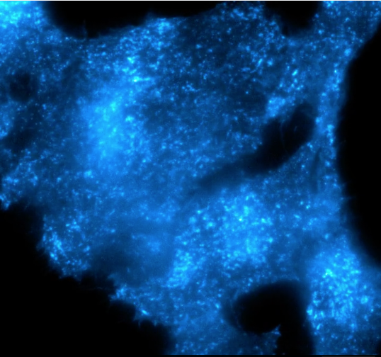
Microscopy encompasses a broader range of imaging techniques used for larger-scale objects, while nanoscopy specifically focuses on achieving high-resolution imaging at the nanometer scale

Focus on your research

Abbelight masters the whole spectrum of fluorescence microscopy while pushing back the limits of higher-throughput imaging techniques : Widefield and Total Internal Reflection Fluorescence (TIRF) microscopy

  • Widefield
  • TIRF
Abbelight pushes the limits of throughput even further with its innovations. ASTER, a patented uniform illumination and Smart multi-color detection strategy gives access to:
Uniform, speckle-free, largest field of view
Simultaneous multicolor microscopy imaging without chromatic aberration up to 4 colors
Adaptable, automatic, quantitative and reproducible TIRF microscopy to define the optimum TIRF angle position compatible with the combination of microscope, objectives and accessories

Abbelight™ ASTER

The largest, homogeneous field of view on the market

The patented Abbelight Adaptative Scanning for Tunable Excitation Rendering (ASTER) technology is a unique, versatile illumination scheme that creates the largest uniform field of view (FOV) of a sample on a multi scalable platform.

• 150×150 µm² field of view
• Illumination adaptable to the sample
• TIRF, HiLo or EPI illumination modes
• More quantitative data
• Homogeneous illumination, no interference patterns in the image

Learn more

Microscopy multicolor imaging

An easy and reliable method for imaging up to 4 biological structures simultaneously

Robust optical design and software architecture leads to highly stable sequential or simultaneous multicolor imaging without chromatic aberration, optical artefact, or misalignment over time.

• Dual camera and four channels: save time and image up to 4 structures simultaneously
• Easy and precise multi-channel digital alignment to recalibrate the channels and correct alignment faults with < 0.7 pixel precision of alignment (no full stop)

Learn more